Iota subfamliy
Protein kinase C is an effector in the G protein-coupled receptor system. It is water-soluble in an inactive state, and is freely present in the cytosol. It becomes a membrane-bound enzyme after activation. The activation of protein kinase C is lipid-dependent and requires the presence of membrane lipid DAG, which is also Ca2+-dependent and requires an increase in the Ca2+ concentration in the cytosol. When DAG appears in the plasma membrane, protein kinase C in the cytosol is bound to the plasma membrane, and then activated by Ca2+. Like protein kinase A, protein kinase C belongs to multifunctional serine and threonine kinases.
Activation
The activity of PKC depends on the presence of calcium ions and phospholipids, but only in the presence of diacylglycerol (DAG), an intermediate product of phospholipid metabolism, calcium ions at physiological concentrations work, because DAG can increase the affinity of PKC for substrate. Phosphatidylinositol-4,5-bisphosphate (PIP2) is hydrolyzed by phospholipase-C to generate DAG and IP3. IP3 promotes the release of intracellular calcium ions and acts synergistically with DAG in the activation of PKC. 12-o-tertradecanoylphordol-13-acetate (TPA; or phorbol-12-myristate-13-acetate, PMA) is a tumor-promoting agent, because its base structure is similar to DAG Simulate DAG at concentration, activate PKC, and increase PKC affinity to 10-7M. PKC is the receptor of TPA. When TPA is inserted into the cell membrane, it can directly activate PKC instead of DAG. When cells are treated with high doses of TPA, PKC in the target cells can be rapidly depleted, which in turn affects cell signaling. A variety of chemicals or antibiotics have an inhibitory effect on PKC activity. According to the different PKC target sites of inhibitors, inhibitors can be divided into two groups: one group is inhibitors that act on the catalytic region, and they can be conserved with protein kinases. Residues bind, so there is no obvious selectivity for PKC; the other group is inhibitors acting on the regulatory region, which can be combined with Ca2+, phospholipids and diacylglycerol/phorbol esters, and thus have higher selectivity.
Iota subfamliy
Protein kinase C iota type is an enzyme that in humans is encoded by the PRKCI gene. PKC-ι is a member of the PKC family located in chromosome 3 at 3q26.2, and is a human oncogene. PKCs can be overexpressed in various types of cancer, including ovarian, lung, head and neck cancer, and PC. Elevated levels of PKC-ι have been correlated with poorer prognosis. It was found that patients with lung cancer who had elevated levels of PKC-ι during the early stages were 10 times more likely to succumb to mortality from the disease, compared with those who had low levels of PKC-ι. PKC-ι is also involved in several oncogenic signaling path- ways. For these reasons, the in vitro effects of two novel aPKC inhibitors, 5-amino-1-(1R,2S,3S,4R)-2,3-dihydroxy-4-methylcyclopentyl)-1H-imidazole-4-carboxamide (ICA-1) and 2-acetyl-1,3-cyclopentanedione (ACPD), on the normal RWPE-1 cell line, and the DU-145 and PC-3 PC cell lines, were investigated in the present study. ICA-1 has been shown to target PKC-ι, whereas ACPD has been shown to target PKC-ι and PKC-ζ.
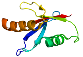 Figure 1. Protein structure of Protein kinase C iota.
References
- Micol V; et al. Correlation between protein kinase C alpha activity and membrane phase behavior. Biophysical Journal. 1999, 76 (2): 916-27.
- Steinberg SF; et al. Distinctive activation mechanisms and functions for protein kinase Cdeha. Bioehem J, 2004, 384 (Pt 3): 449-459